
A vaginal ultrasound, also called a transvaginal ultrasound, is a type of medical imaging that looks for abnormalities inside your pelvis. This includes the vagina, uterus, ovaries, fallopian tubes, bladder, and surrounding areas.
The procedure involves inserting a small probe into the vagina and gently moving the tool around during the test. Most people don’t find a vaginal ultrasound painful, but it can cause discomfort.
A healthcare provider might recommend a vaginal ultrasound to investigate symptoms like pelvic pain or bleeding or to help prepare for surgery. The imaging can show how thick the endometrium (lining of the uterus) is and whether there are cysts, uterine or ovarian tumors, or anything else of concern.
You might need a vaginal ultrasound for the following:
- Ectopic pregnancy (pregnancy occurring outside the uterus)
- Unexplained pelvic pain
- Abnormal vaginal bleeding
- Infertility
- Differences in the uterus or ovaries present since birth
- Suspected endometriosis (when endometrial, or uterine, tissue grows outside of your uterus)
- Suspected adenomyosis (when endometrial tissue grows into the uterine walls)
Preparation for a vaginal ultrasound is minimal—you can eat and take your usual medications unless advised otherwise. Your provider may request an empty or partially filled bladder for better imaging, so ask about specific instructions.
For comfort, consider bringing a loved one or requesting a chaperone. Bring your ID and insurance card, and confirm insurance coverage beforehand. Before the procedure, you’ll privately undress and be provided a gown or sheet for coverage.
A vaginal ultrasound works similarly to other types of ultrasound that you might be more familiar with. The difference here is that the probe goes inside of you.
An ultrasound wand called a transducer is inserted into your vagina. It creates sound waves, which bounce off the organs and tissues in your pelvis. These results are sent to a computer, which creates a two-dimensional gray-scale picture of your insides called a sonogram. Depending on the exam room setup, you might be able to see the sonogram during the procedure.
The following healthcare providers may perform a vaginal ultrasound:
- Sonographer: A technician trained in performing ultrasounds
- Radiologist: A medical doctor who specializes in diagnostic imaging, including ultrasounds
- Gynecologist: A medical doctor who specializes in the female reproductive system
- Obstetrician: A medical doctor who specializes in the care of pregnant people and the delivery of babies
During the Test
Here’s what you can expect during your appointment:
- Preparation: You’ll be asked to lie flat on a table with your knees bent and feet elevated, sometimes in stirrups. A gown, sheet, or paper will cover your abdomen.
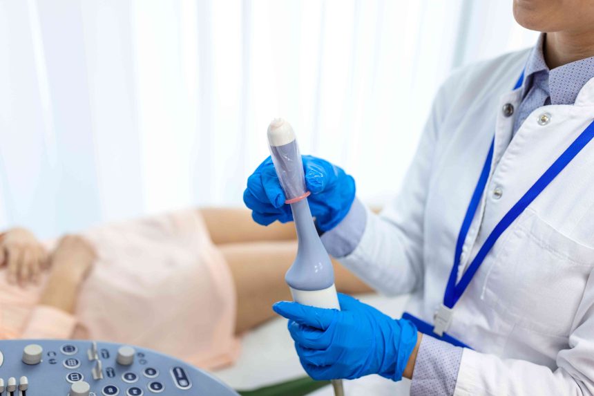
- Insertion of the ultrasound wand: The healthcare provider will insert a small ultrasound wand (about 2-3 inches long) into your vagina. The wand is smaller than a standard pelvic exam speculum and will be covered with a condom for sanitation and lubricant gel for easier insertion.
- Self-insertion option: If it makes you more comfortable, ask your provider if you can insert the wand yourself. Some providers offer this option.
- Imaging process: The provider will gently move the wand to capture images of your internal organs.
- Duration of the procedure: The ultrasound typically lasts 15-45 minutes.
- Expected sensation: While the procedure is usually not painful, some people may feel a bit of discomfort during the exam.
After the Test
Once the ultrasound is over, the wand will be gently removed. You should be given some time to privately clean up and get dressed.
This is an outpatient procedure, meaning you can go home afterward. There shouldn’t be any lasting pain or symptoms.
Vaginal ultrasound is a safe procedure with no radiation exposure.
Some people—particularly those with a history of sexual trauma—might find it emotionally distressing due to its intimate nature. Consider disclosing this to your healthcare provider in advance for increased sensitivity to your situation.
People with vaginismus (a condition where you experience pain during penetration) or other sources of pelvic pain may find the imaging procedure uncomfortable or painful. If you are concerned, talk to your provider about pain management before the procedure.
Because a condom is put over the ultrasound wand and lubrication is put onto the wand, you should let your provider know beforehand if you have any allergies to either item, such as a latex allergy.
If you are unable to have a vaginal ultrasound due to vaginismus or another reason, your provider may recommend an abdominal ultrasound of the pelvis instead.
Once your exam is complete, the ultrasound images will be sent to a radiologist for interpretation. The radiologist will send your provider a copy of the images with their notes. If a radiologist performed your vaginal ultrasound, they may discuss their findings with you after the exam.
Depending on the results, you may need to schedule a follow-up appointment or exam with your healthcare provider.
Interpreting Your Results
The healthcare providers who perform vaginal ultrasounds and study the images are trained to spot abnormalities that might include:
- Pelvic, ovarian, or uterine masses
- Thickened endometrium
- Enlarged uterus
Those findings might indicate certain diagnoses, such as:
- Cancers of the uterus, vagina, ovaries, and other pelvic organs
- Ovarian cysts or fibroids
- Endometriosis or adenomyosis
- Ovarian torsion (twisting ovaries)
- Ectopic pregnancy
- Infections
A vaginal ultrasound doesn’t treat any of these conditions, but it can help your healthcare provider decide the next steps for further diagnostic procedures or treatments.
Vaginal ultrasound is an internal form of ultrasound where a wand is inserted into the vagina. Sound waves create an image of the internal pelvic structures, including the uterus, vagina, fallopian tubes, and ovaries.
The procedure is not painful for most people but may cause discomfort. A vaginal ultrasound can help guide diagnosis or treatment for several gynecological conditions, such as cancers, endometriosis, and ectopic pregnancy.